0
£0.00
0 items

A new generation of machines is emerging. Using the latest technology, it provides exquisite structural detail and performs better in visualising those challenging and deep structures.
I recently reviewed the GE LOGIQe, S7, S8 and E9 next to each other in the same session and concluded that the reputation of the E9 as the workhorse and standard in MSK Ultrasound was well deserved, despite many other manufacturers producing increasingly fierce competition.
So, now there is the E10. GE kept the same MSK probe, similar clear keyboard layout, nice big control touchscreen and very clear monitor. But what about the imaging? I took it for an extensive test drive, with the help of Rick Miles, GE Healthcare Lead Clinical Application Specialist POC Ultrasound, specifically looking for the MSK application in the traditionally more challenging areas.
| Conclusion, in short: It has raised the standard in MSK ultrasound imaging. It makes scanning very easy and lifts visualising the structural detail to a new level. |
The E9 does a very good job visualising superficial structures, but the E10 makes it that much easier to get a good image. Combined with excellent Doppler and shearwave elastography it performs very well when assessing tendons, ligaments and joint capsula. For the deep structures, especially hamstring and adductor muscles it shows great detail, but needs some tuning in to be able to confidently assess this. In my view, for MSK imaging there should always be a pre-set for deep muscles that works well. And as far as identifying and tracing the peripheral nerve branches, that is so much easier, with no need to adjust settings to optimise the image. Just scan.
This new generation of machines will provide the clinician with new details and insight that will question some of our current beliefs in MSK injury management. If the E9 is a turbocharged sports car, the E10 is a supercar that just needs some tuning to perform at its best in all circumstances.
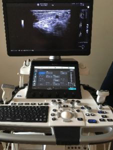
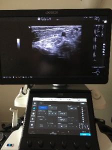
The GE Healtcare LOGIQ E10
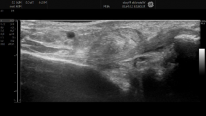
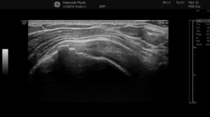
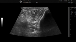
Testing included posteromedial knee (visualising capsula, meniscus and semimembranosus at the same time), lateral elbow, anterior and lateral hip, hamstrings, adductors, calf muscles and Achilles tendon complex, tibial nerve branches distal to the tarsal tunnel, lateral ankle, rotator cuff interval (tendons, ligaments and rotator cuff cable), posterior shoulder joint.
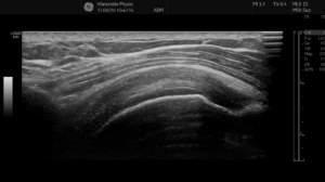
CFL and the fibular attachment
Supraspinatus in longitudinal plane
Rotator cuff cable in transverse and longitudinal plane
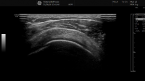
Posteromedial knee joint: capsula, meniscus and semimembranosus tendon
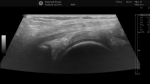
Lateral elbow
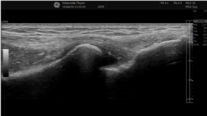
Anterior compartment