0
£0.00
0 items
If you’ve been in the ultrasound game a while, you’ll no doubt be familiar with quite a few makes and models of various systems – particularly if you’ve been to one of our courses or read our blog.
GE, Sonosite, Mindray, Clarius and Sonoscape and Alpinion are names that likely ring a bell even if you haven’t scanned on them.
At SMUG we are very biased towards GE! All SMUG tutors use GE in their clinics, and we have all been using them for many years. We also partner with GE for all our courses, and they provide all the high-end machines for our courses. TBH we wouldn’t have it any other way! GE have a full range of ultrasound machines, for beginners to experts, from big budgets to smaller budgets! Their machines start with the GE V Scan Air handheld machine (for under £5000) to their top of the range cart-based machine, the E10 (for over £50,000!).
However, we also like to try other machines. There is one brand you may not have come across and that doesn’t often get a mention when it comes to MSK ultrasound in our practice. It’s Canon – yes, the company that makes cameras and printers.
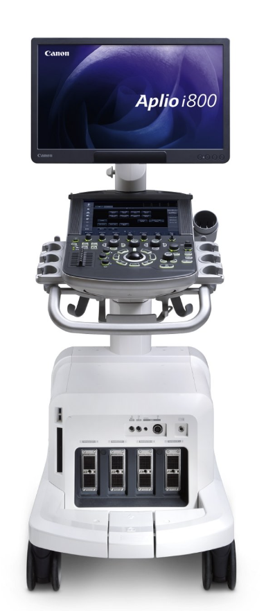
Canon Medical bought Toshiba in 2016 so they also make medical imaging equipment including ultrasound scanners and I must say, they’re impressive. I had the good fortune to use their i800 Prism edition for a couple weeks in my clinic recently so thought I’d post a review of my experience here.
The Canon was very easy to use considering how many buttons are on the console and how much is happening on the touchscreen and monitor. Instead of being overwhelming, it was very intuitive. Finding depth, focus, gain and frequency on most systems is reasonably straight-forward but what surprised me was that in addition to the basics, I could review, edit and restart studies with ease. It just all made sense and I wasn’t left staring at the screen for long before figuring it out.
The machine’s image quality is very, very impressive! Let’s be clear this is not a cheap machine; in fact, it is one of the most expensive of the market.
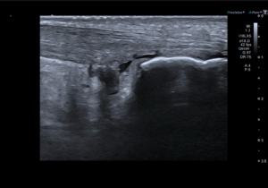
Longitudinal image of Achilles tendon showing a retrocalcaneal bursitis and distal tendinopathy with a partial ventral tear. Video:
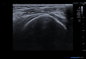
The presets on the high-frequency linear probe barely needed any tweaking despite scanning a range of patient shapes and sizes. I’d occasionally adjust the frequency but nothing else. I scan quite
a few hips in my day-to-day work, aswell as lateral hip tendons and hamstring origins. These can be a tough area to confidently visualise. I was impressed that the Canon could demonstrate these so
clearly. The needle visualisation at these depths was also very good (see below)
Video 1: Ultrasound guided injection for ITB friction syndrome. The injectate flows under the ITB.
Video 2: Ultrasound guided injection hip joint injection.
The Canon has a different image appearance/texture to the GE – it is slightly smoother - but it does this without losing any contrast resolution so differentiating between structures is easy as boundaries remain clear. As I said I am bias to GE, so I personally prefer the E10 image but that’s probably because that is what I am used to. Of course, ultrasound is user dependent, and one must always be conscious of anisotropy, but I felt the Canon dampened the effects of anisotropy, like you
would expect of a high-end machine.
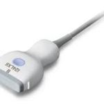
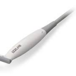
Canon’s high-frequency linear (4-18MHz) is a very versatile probe and was suitable for most of my needs. They have a small curvilinear probe with a frequency range from 2-10MHz which I didn’t use
much as the linear performed so well. However, I like the idea of this small convex probe. The small footprint and higher frequency made it feel more like a linear probe and was easier to handle than the traditional larger convex probes.
|
|
|
|
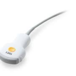
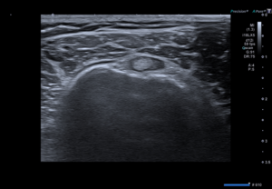
I had a run of Morton’s neuromas in the weeks I had this system and scanning with the 22MHz ‘hockey stick’ was amazing. Not only was the image really clear, but the slim footprint made access to small spaces very easy, and the ergonomic shape of the probe felt nice to handle. The spatial resolution possible with the ‘hockey stick’ was impressive. I was able to easily appreciate internal fascicles of small nerves like the superficial peroneal nerve.
I believe Canon also have a 24MHz and a 33MHz linear probe, but I didn’t test it out on this trial.
As mentioned earlier, Canon’s imaging is very good. Of note for MSK, their panoramic view is easy to use and renders exquisite detail. They also have a horizontal split screen function which is particularly useful if you want to compare elongated structures such as finger tendons or the Achilles tendon with an asymptomatic contralateral side. The traditional vertical split screen option is still available if needed. To be honest, for a high-end machine it has all the “add ons” you would expect.
They aslo have advanced shearwave technology and although it’s used widely in hepatology and breast imaging, the jury is still out for MSK, so I didn’t spend any time with it on this occasion.
Canon’s SMI Doppler technology is a welcome edition. SMI stands for ‘Superb Microvascular Imaging’ and is unique to their systems in the way it can detect and display slow flow and micro vessels. I see a lot of tendons and a lot of tendinopathy, and I know the amount of vascularity I can detect varies significantly depending on the system I’m using. Without a doubt, I was appreciating more vascularity with Canon’s SMI than ever before. This was clear in the rotator cuff and especially around the coracohumeral ligament and the rotator interval. It’s interesting to ponder how much flow is present in symptomatic patients even when we can’t detect it on some of our systems. More research in the area is required for us to fully understand the implications for practice, but I am sure there will be many!
It can’t all be sunshine and rainbows – there are some disadvantages to this system that need to be considered. This unit is not a point-of-care system so the footprint may be larger than a small clinic can accommodate (approximately 63 x 97cm at the base). In places like London, where every square foot in a clinic is priced at a premium, you’ll need to make sure your room is big enough to start with and then consider if this kit is worth that precious real estate.
It’s not portable, although it’s on wheels! It has a good turning circle and is adjustable for ergonomic reasons, it’s not really designed to be moved from room-to-room. At 115kg it’s hefty and is not easy to simply move from room to room on a regular basis. This system would be best set up in a single room.
It’s expensive… but I can see that you get what you pay for. Yes, this system is more expensive than many units on the market but the technology out performs the cheaper ones by far. It’s great if you need to see deeper structures, such as the hip joints, lateral hip tendons and soleus for example. The Canon would be a massive investment, but the unit would not need to be replaced for a long time.
There’s a lot to say about my entre into scanning on a Canon. The Pros include excllent image quality, intuitive and comfortable use, and advanced technology which all cumulates in increased diagnostic confidence. The Cons include the relatively high price and the increased size compared to more portable/ point-of-care systems. It won’t be the system for everyone reading this.
If you have any questions or would like further information about machines, please email chris@ultrasoundtraining.co.uk