0
£0.00
0 items

What ultrasound probe should I buy?
For those new to diagnostic ultrasound there are many questions you need to try and answer. On the SMUG website we have tried to answer some of these questions; these vary from how to get started in MSK ultrasound, what machine to buy and how much to spend, how to become competent in MSK Ultrasound and how to make your money back (or not!).

Getting the right information for your specific needs and requirements can be quite tricky. Over the years we have written many blogs that will help you (see image above). One question we often get asked on our courses and via email iswhat probe do I need to buy musculoskeletal ultrasound and how many do I need? We thought we would try and answer this and introduce some of the considerations in relation to probes for MSK ultrasound.
Integrating diagnostic ultrasound is not a cheap option. In our opinion the benefits for you and your patients outweigh the costs. However, many people do waste money on probes that they do not necessarily need and also on machines that will not perform well enough or have a good enough image quality for musculoskeletal ultrasound.
When it comes to ultrasound equipment it is important you do not buy something because it is the cheapest (“buy cheap, buy twice” springs to mind!). Inadequate equipment with poor image quality is going to lead to mis-diagnosis and make your job much harder. Diagnostic ultrasound is an operator and machine dependent modality. It does not matter how good you are at scanning, if you have poor equipment you will not produce a high quality image.
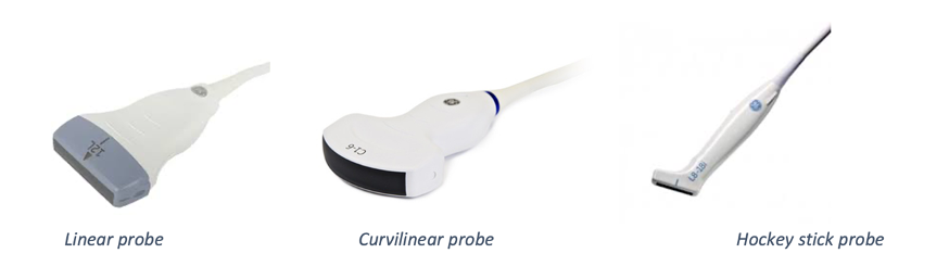
Here are the three probes used in MSK ultrasound.
There are several elements to consider regarding the above probes:
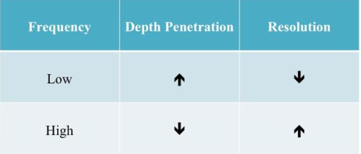
1. Frequency of probe –This relates to the frequency of the pulse wave it is capable of emitting. The probes, nowadays, are able to emit a range of frequencies – this is called the bandwidth. For example, a 7–15 MHz probe has a bandwidth from 7 to 15 MHZ.
Basically, the lower the frequency, the deeper the pulse wave can penetrate and return, though at the expense of image quality. Therefore, these probes are used to image deeper structures such as those around hip joint. The higher frequency probes have better image resolution (spatial resolution), whilst sacrificing depth of penetration. Higher frequency probes are used for imaging more superficial structures such as the Achilles tendon.
High frequency probes also need a machine with the adequate processing power to produce a clear image with good spatial resolution. Some machines actually have a higher frequency probe but have a poorer image quality to a lesser frequency probe. Remember, a high frequency probe optimises the structures in the first few centimeters of the image only, not all the image. It is always worth trying any machine/probe combo before buying them. Try before you buy!
For MSK scanning you will require a relatively high frequency probe to ensure adequate image quality. However, frequency is just one component that influences image quality. I would suggest that for MSK imaging the probe needs to go up to a minimum of 12MHz. Therefore, a 3-12 or 5-12 MHZ probe is what you are after. All modern machines are likely to have this specification. However, if you are buying an older, second hand machine it is worth checking the frequency of the probe.
2. Footprint – this is the part of the ultrasound probe that interfaces/touches the patient. This is where the ultrasound pulse waves are emitted and returning echoes are received. Depending on type of the probe, the dimensions, area and the shape of the footprint differ. The curvilinear probe has a curved and wide footprint. Whereas the linear probe, this includes the hockey stick probe, flat and thinner. The size of the footprint also varies between manufactures.
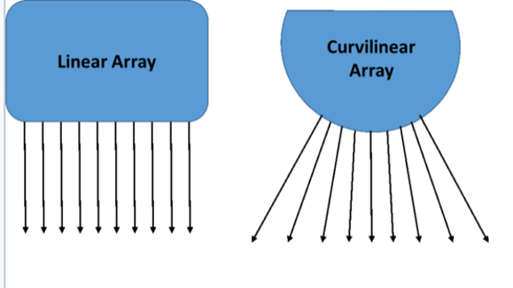
3. Field of view (FOV) – this is the field of vision the probe displays on the screen. The linear and curvilinear both have a wide FOV, which is useful for MSK scanning. The linear FOV has parallel perpendicular lines, in the shape of a square/rectangle. Whereas the curvilinear with a wider depth of field, to improve visualisation of deeper structures.
We will now discuss the basic features and considerations of each MSK probe.
Linear probe
Higher-frequency linear probes (7–15 MHz) provide excellent resolution and image quality for a majority of MSK anatomical structures. Linear probes do lack the penetration depth of lower frequency probes e.g., the curvilinear probe (see below). These probes have a medium sized footprint and so can be manoeuvred successfully around smaller joints and structures.
If you are getting started a high frequency ‘linear’ probe is the key one to buy. You do not need more than one probe to get started. The linear probe is a good all-rounder on most ultrasound machines and can be used to scan all joints. The image will be compromised scanning a deeper structure e.g., scanning a tendon at 2cm versus 6 cm, but it will still produce an adequate image quality.
In my London clinics, we use a GE Loqiq-e and an Alpinion i-cube 7 (both portable machines). Both have just one probe, the GE Logiq-e has the 12L-RS probe (5-13Hz) and the Alpinion L3-12T (3-12MHz). Both probes are more than adequate for MSK. We also use these probes for all our ultrasound guided injections in the clinic, from superficial injections such as subacromial bursa or trigger finger injections to deeper injections around the hip such as the proximal hamstring and hip joint injections. Furthermore, both of these machines have a ‘virtual convex’ function, a very useful feature. This ‘virtually’ turns the linear probe into a curvilinear or curved probe. This can further improve visualisation of the deeper structures.
Curvilinear (convex) probes
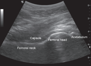
These are normally lower frequency (1-6MHz) to target deeper structures. In MSK scanning these probes are commonly used for scanning the hip and shoulder, particularly in obese or very muscular patients. These allow deeper ultrasound penetration which comes at a cost of poorer resolution. These probes are used by some practitioners for deeper ultrasound guided injections such as the hip and shoulder. However, it is not essential to have a curvilinear probe to carry out these injections.
Hockey stick probe
Hockey stick probes are higher frequency (8-18MHz) and have a smaller footprint. They allow improved probe contact between small irregular surfaces such as when scanning hands and feet. The smaller footprint allows improved probe contact and visualisation. Carrying out ultrasound guided injections into smaller joints such as the wrist & hand and foot joints can be made a little easier using a hockey stock probe as it has a smaller footprint. However, it is not essential to have a hockey stick probe to carry out these injections. Many rheumatologists use hockey stick probes.
It is important to note you can image almost anything with any probe depending on what you are trying to scan. However, usually one probe is more ideal than another, but suppose you have no access to it you can adapt with the other probes if possible.
5. Your job!
The probe that you require will depend on the type of work that you do. For example, we recently had an orthopaedic surgeon attend our ‘cadaveric 2 day ultrasound guided injection course’. His objective from the course was to carry out ultrasound guided hip injections instead of using fluoroscopy guidance. For this he may prefer a linear or curvilinear probe. Whereas another delegate on the cadaveric course was a consultant rheumatologist. He carries out ultrasound scans of the hands and feet, to screen for inflammatory conditions and ultrasound guided procedures for synovitic joints, predominantly smaller joints such as PIP and DIP joints. For this he may prefer a hockey stick probe, which is very high frequency probe with a small footprint, with excellent power Doppler.
Conclusion
As mentioned earlier for most clinicians with a varied MSK caseload, one high frequency linear (up to 12MHz) will be sufficient to get started. Having more than one probe, infact having all three MSK probes (linear, curvilinear and hockey stick probe) would be ideal, but may be considered a bit of a luxury particularly when you get started. As you get more experienced at scanning and are more aware of what is a ‘good’ image versus a ‘great’ image having different probes becomes more important.
If you have any questions about probes or ultrasound machines, then please do not hesitate to contact chris@ultrasoundtraining.co.uk